Whole slide imaging is no longer a technology looking for a use case. It is the backbone of modern pathology, whether a pathologist is signing out a colon biopsy for a patient waiting on results, or a research team is training the next generation of AI models on thousands of annotated slides.
But the requirements for these two contexts are fundamentally different. Understanding those differences matters when choosing a scanner, because the wrong fit costs a lab time, money, and confidence in the images it produces.
When a scanner is used for primary diagnosis, its output replaces what the pathologist would otherwise see under a microscope. That changes everything about how the device is evaluated.
Regulatory standing comes first. In the EU, a scanner used for primary diagnosis must carry CE IVDR approval, confirming it meets the performance, safety, and quality standards required under the In Vitro Diagnostic Regulation. In the United States, the regulatory path runs through the FDA. Until a scanner has FDA clearance, US labs looking to use digital pathology for primary reads must independently validate the system under their CLIA certification. This is a structured, evidence-based process where the lab demonstrates that diagnoses made on digital images are concordant with those made on glass.
Image quality is non-negotiable. Dermatopathology specimens with subtle melanocytic features, GI biopsies where glandular architecture drives the diagnosis, soft tissue cases that depend on cellular morphology at high magnification: these are the everyday workload of a clinical lab. The scanner must handle them without compromise in color fidelity, sharpness, or consistency across slides.
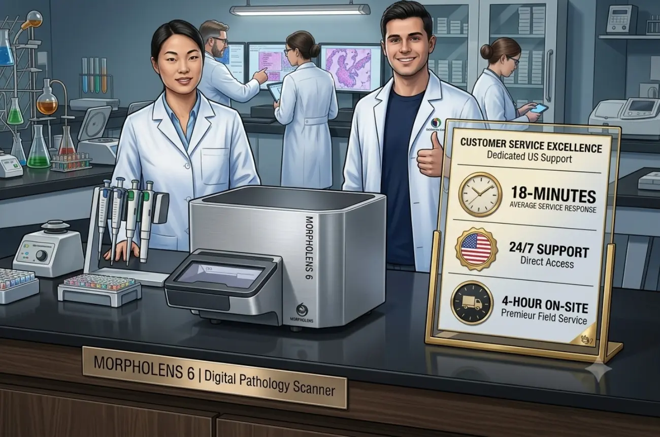
Reliability and uptime matter as much as image quality. A clinical lab cannot afford multi-day downtime waiting for a service engineer. Scanners in this setting need to run continuously, handle mixed slide loads, and be supported by a responsive service infrastructure.
Integration is expected, not optional. The scanned images need to flow into the lab's LIS/LIMS and digital pathology viewing platform without manual workarounds. Enterprise deployments demand this.
Research labs operate under different constraints, but that does not mean their requirements are lower. They are simply different.
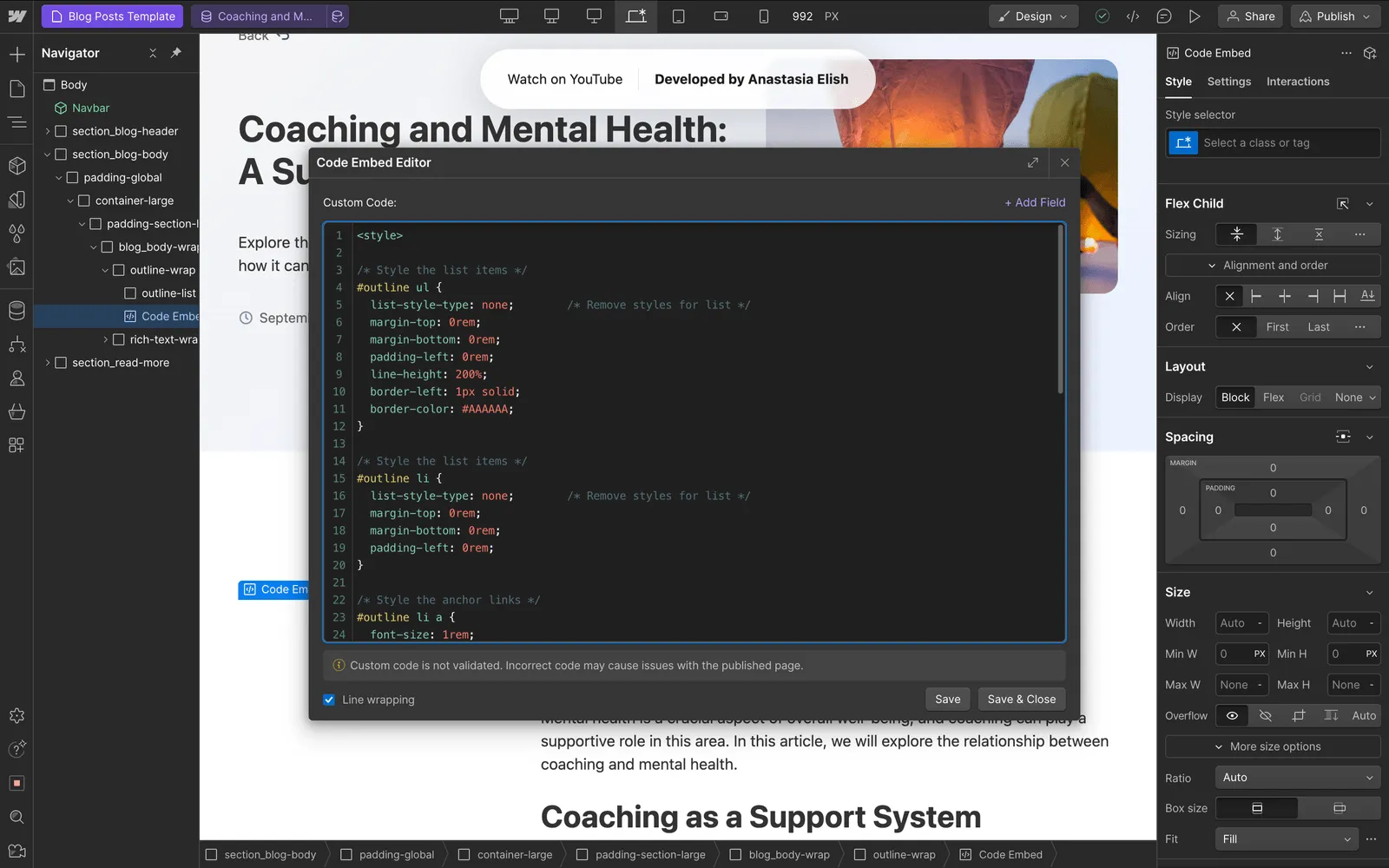
Throughput and flexibility take priority. A research lab studying tumor microenvironments might scan 500 slides in a week, then shift to fluorescence work, then return to brightfield H&E. The scanner needs to accommodate varied protocols, tissue types, and scan settings without requiring recalibration for each run.
Data quality must be reproducible. Research findings that depend on image analysis, whether manual scoring or algorithmic quantification, require that the scanner produce consistent results across batches, across weeks, across operators. Drift or variability in scan quality introduces noise that can undermine a study.
Cost structure matters. Research budgets are finite and often grant-funded. A scanner that delivers clinical-grade image quality without the price tag of systems designed primarily for high-volume hospital deployments is a meaningful advantage.
Open data access is valued. Researchers want standard file formats, accessible APIs, and the ability to export and process their data freely. Proprietary lock-in is a friction point in academic settings.
Morphle scanners were built to perform in both of these environments, and multiple hundreds of deployed units across India and the United States prove it in practice.
On the clinical side, Morphle scanners carry CE IVDR approval, meeting the regulatory bar for primary diagnosis in the European Union. In the United States, multiple labs have successfully completed their CLIA validation studies on Morphle systems, putting them into active clinical use for primary reads. Morphle is also on the path toward FDA clearance for its scanners, a process that will further expand access for US labs looking for a regulatory-cleared digital pathology solution.
The clinical specialties where Morphle has gained the strongest traction reflect the areas where image quality is most heavily scrutinized: dermatopathology, GI and colon biopsies, and soft tissue pathology. These are the subspecialties where pathologists will immediately notice if a scanner falls short, and Morphle's adoption in hundreds of labs across these disciplines speaks to the confidence users have in the platform.
On the research side, Morphle systems are deployed across academic institutions running high-volume scanning for tissue studies, AI training datasets, and multi-site collaborations. The same image quality that satisfies clinical pathologists gives researchers the consistency they need for quantitative analysis and reproducible results.
Morphle is also used within larger enterprise-scale digital pathology deployments, where reliability, workflow integration, and scalability are critical requirements. This means not just a single scanner on a benchtop, but multi-unit installations integrated into lab workflows with centralized storage, network-based image access, and the service infrastructure to keep everything running. LIS integration is part of this picture, and a non-negotiable for any lab moving primary diagnosis onto a digital platform.
For enterprise-grade deployments, Morphle has proven to be a popular choice on the strength of a specific combination of factors:
Whether a lab is scanning 10,000+ slides a week for clinical sign-out across multiple units, or 50 a day for a research study, the platform scales to match.
The distinction between clinical and research use cases is real, and it shapes how labs evaluate scanners. But the best outcome for any lab is a platform that does not force a choice between the two. A scanner validated for primary diagnosis and trusted in demanding subspecialties like derm and GI, while also flexible and cost-effective enough for high-throughput research, removes the need to maintain separate imaging infrastructure for separate purposes.
That is what Morphle was designed to deliver, and what hundreds of labs worldwide are already using it for.
To explore how Morphle can fit your unique use case, Contact our team.

.webp)
.webp)
.webp)
.webp)
.webp)
.webp)