Whole Slide Imaging (WSI) and tele-pathology are no longer separate disciplines; they are two sides of the same coin. A high-performance digital pathology scanner converts glass slides into richly detailed digital files that pathologists can review from anywhere in the world. This blog breaks down how the two technologies integrate, why it matters for diagnostics, and what to look for when choosing the right scanning solution.
Whole Slide Imaging (WSI) refers to the process of digitizing an entire glass slide at high resolution using an automated microscope slide scanner. The output, a whole slide image, replicates the optical fidelity of a conventional microscope, but in a shareable, zoomable digital format.
Telepathology is the transmission of pathological data, images, annotations, reports, over electronic networks for the purpose of remote diagnosis, second opinions, or education
Historically, telepathology relied on static JPEG captures or real-time robotic microscopy. WSI changed the equation entirely. Instead of sending a single field of view, a digital pathology scanner now captures the full tissue section, giving the remote pathologist complete contextual visibility, the same view they would have at the bench.
The integrated WSI-telepathology workflow follows a clear sequence:
This pipeline compresses what once required a physical courier service for slides into a process that can be completed in minutes.
Not all scanning systems deliver equal results. These are the parameters that matter most:
Optical resolution determines the finest detail that can be resolved. Most diagnostic workflows require 0.25 µm/pixel (40× equivalent) for cytology and high-detail morphology, and 0.5 µm/pixel (20× equivalent) for routine histology.
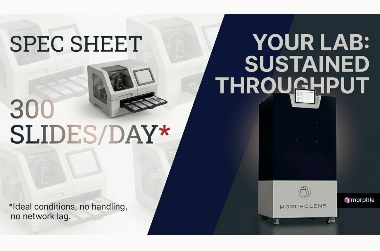
Scan speed directly impacts lab throughput. Enterprise labs processing hundreds of slides daily need scanners that can complete a standard slide in under 90 seconds.
Focus accuracy — both pre-scan focus mapping and real-time z-stack adjustment- is essential for uneven tissue sections common in slide scanner histology applications.
File format and compression affect storage costs and image fidelity. Formats such as SVS, NDPI, and TIFF are industry standards; lossless compression preserves diagnostic accuracy.
Connectivity and integration — compatibility with DICOM standards, LIS/PACS integration, and API access are non-negotiables for seamless telepathology deployment.
Frozen section consultation — Intraoperative slides are scanned and transmitted to an off-site pathologist for real-time surgical guidance, eliminating the need for on-call pathologists at every surgical facility.
Second opinion and tumor boards — Complex or rare cases benefit from multi-institutional input. WSI enables seamless sharing without the risk of slide loss or degradation.
Histology research and education — Universities and research centers use whole slide scanning to build digital teaching archives. Students navigate real diagnostic cases at full resolution, regardless of location.
Low-resource and developing-market diagnostics — Portable and compact digital pathology scanners are bringing specialist-level diagnosis to underserved regions, a trend discussed extensively in industry conversations around deploying "digital pathology in a suitcase" to expand the geographic reach of telepathology.
Oncology and pharmaceutical trials — Centralized pathology review for multi-site clinical trials relies on WSI to standardize tissue evaluation across geographies.
If your lab is evaluating a pathology slide scanner, here is a practical checklist:
The WSI-telepathology ecosystem is evolving rapidly. Several developments will shape the next decade:
AI-assisted diagnosis — Computational pathology algorithms are being embedded directly into scanning workflows, flagging abnormalities, quantifying biomarkers, and triaging cases before a pathologist reviews them.
Federated learning — Labs are training AI models on distributed datasets without centralizing patient data, addressing privacy constraints while building powerful diagnostic tools.
Portable and compact scanners — The next frontier is bringing whole slide imaging capability to settings that could never support conventional lab infrastructure, point-of-care, field hospitals, and remote diagnostic centers.
Multimodal integration — Correlating WSI data with genomics, radiology, and clinical records is moving pathology toward truly integrated precision medicine.
Among the digital pathology companies building for this future, Morphle Labs stands out for its focus on high-throughput, high-fidelity scanning combined with intelligent workflow integration. Morphle's digital pathology scanner is engineered to deliver the resolution, speed, and connectivity that modern telepathology demands, whether you are running a busy diagnostic lab, supporting a multi-site clinical trial, or building an AI training dataset.
Morphle Labs understands that the value of whole slide imaging is only realized when the image gets to the right pathologist at the right time. That's why their platform is built with seamless remote access, LIS integration, and DICOM compliance at its core, not as an afterthought.
For labs assessing histology scanner options, Morphle Labs offers a solution designed to fit demanding diagnostic environments without compromising on image quality or operational simplicity.
Whole Slide Imaging and telepathology are not parallel technologies; they are a single, integrated capability. WSI provides the high-fidelity digital substrate; telepathology provides the distribution layer. Together, they dissolve the geographic constraints that have historically limited access to expert pathology.
The lab that invests in the right digital pathology scanner today is not just buying hardware; it is buying the ability to deliver expert-level diagnosis anywhere, collaborate instantly, and deploy AI diagnostics as the field matures.
Talk to the Morphle Labs team today. Whether you are exploring your first digital pathology scanner or upgrading an existing workflow, our specialists will walk you through a personalized demo and help you identify the right configuration for your lab's volume, use case, and compliance requirements.
👉 Request a Demo with Morphle Labs; and take the first step toward a fully connected, AI-ready pathology workflow.

.webp)
.webp)
.webp)
.webp)
.webp)
.webp)