Cytology presents unique imaging challenges; three-dimensional cell clusters, variable thickness, and the need for exceptional fine-detail resolution. A whole slide imaging system designed for cytology requires precise focus, advanced Z-stacking, and workflow-driven best practices. This article outlines technical, operational, and practical guidelines to ensure high-quality digitization of cytology slides using modern scanners and imaging tools.
Whole Slide Imaging System
A combination of an optical scanner, imaging engine, and WSI viewer used to digitize entire cytology slides at diagnostic quality.
Whole Slide Scanning
The process of converting glass cytology slides into high-resolution digital images for review, annotation, or telepathology.
Digital Pathology Slide Scanner / Automated Microscope Slide Scanner
Devices that automate scanning, focusing, Z-stacking, and imaging to ensure accurate digital representation of complex specimens.
Pathology Slide Scanner / Histology Scanner / Microscope Scanner
General terms used for scanners that digitize histopathology and cytology specimens.
Cytology Slide Scanners
Specialized scanners capable of capturing multi-plane focus (Z-stacking) to visualize thick cellular clusters.
Cytology samples require careful preparation before scanning. Slides typically undergo:
A modern whole slide imaging system must support this end-to-end workflow reliably and efficiently.
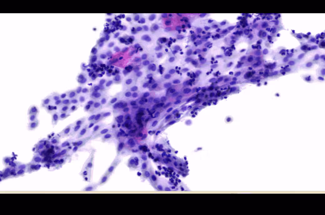
Cytology clusters are inherently three-dimensional. A flat single-plane scan misses nuclear contours, chromatin patterns, and overlapping structures.
High-performance cytology digital pathology workflows rely on variable or automated Z-stacking to recreate depth accurately.
Cytology slides often contain large smear areas requiring extensive scanning. Systems must optimize speed so labs can maintain throughput.
Consistent illumination, accurate color reproduction, and high NA (numerical aperture) optics are essential to ensure diagnostically relevant detail.
Cytology images generate large files—especially with stacked focus layers. Efficient compression algorithms and viewer-side rendering are key for smooth virtual microscopy.
When developing AI in histopathology or cytology, image quality consistency is crucial. AI models require stable acquisition parameters and high-resolution imaging.
Despite these limitations, digital cytology continues to grow rapidly, especially as digital pathology companies address the unique challenges of these slides.
Cytology WSI must align with clinical regulations, often involving:
A compliant whole slide imaging system reduces medico-legal risk by maintaining robust logging and storage integrity.
Remote interpretation allows cyto-pathologists to support satellite centers instantly.
Digital archives allow peer review, inter-observer comparison, and proficiency testing.
Z-stacked datasets are invaluable for feature extraction and training classifiers.
Digital slides support interactive virtual microscopy for cytology students.
Increasingly, labs use cytology WSI for Pap smears, FNAC, and liquid-based cytology evaluations.
Affordable slide scanners are making these applications accessible even for mid-sized laboratories.
When selecting a system, consider the following:
For help choosing the right device, explore: Morphle slide scanners
Systems will intelligently capture more layers in dense clusters and fewer in sparse areas.
AI-based autofocus will reduce scanning time while improving clarity.
As bandwidth improves, more labs will centralize cytology data for telepathology.
Combining cytology WSI with molecular and fluorescence imaging will become more common.
Hybrid models integrating live automation with stored WSI files are emerging.
Morphle’s scanners are engineered to meet cytology’s demands with:
These capabilities help pathology labs achieve consistent results using cost-effective, reliable imaging technology.
Contact our Team for sample cytology scans.

.webp)
.webp)
.webp)
.webp)
.webp)
.webp)