Research institutes are rapidly adopting digital pathology scanners to improve sample reproducibility, accelerate analysis, enable collaboration, and build AI-ready datasets. With high-quality imaging, streamlined workflows, and scalable storage, digital systems are transforming how labs conduct research and validate findings at scale.
For a research-focused example, explore our work with SRMC: Case Study
Digital Pathology Scanners
High-resolution scanners that convert tissue or cell samples into detailed digital slides for analysis, archival, and sharing.
Whole Slide Imaging Scanner
A system that captures entire tissue sections at magnifications suitable for histopathology, enabling zoom-in viewing without loss of detail.
Microscope Slide Scanner
A digital replacement for the light microscope, offering consistent, calibrated images ideal for quantification and research documentation.
WSI Viewer
Software that renders large whole-slide images instantly, allowing researchers to navigate, annotate, and collaborate in real time.
Virtual Microscopy
The digital equivalent of microscopic review, allowing students and researchers to view slides from any device.
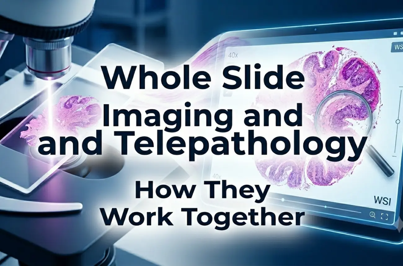
Telepathology
Remote sharing and consultation of digital slides to connect research teams across institutions or continents.
DICOM Pathology
A standardized format for storing and exchanging digital pathology images.
AI in Histopathology
Machine learning tools that analyze patterns, classify structures, or quantify biomarkers—dependent on consistent, high-quality digital slides.
The research workflow today demands speed, scalability, and reproducibility. A Digital Pathology Slide Scanner integrates seamlessly into this environment.
Samples are prepared on glass slides and loaded into the scanner. The system performs tissue detection, auto-focus, image stitching, and color balancing to produce a high-fidelity whole-slide image. Researchers then review the image through a WSI viewer, annotate findings, export regions of interest, and share results across teams.
This workflow supports everything from basic science discovery to translational research, validation studies, and multi-site collaborations. Digital images eliminate variability in microscope optics and ensure that everyone—regardless of location—evaluates the exact same data.
Research often requires sub-cellular clarity. Optical design, sensor quality, and illumination uniformity all influence image fidelity.
Studies relying on stain intensity or biomarker quantification require stable, calibrated imaging that does not drift over time.
Uneven or thick tissue sections require advanced focusing strategies to maintain clarity across the entire specimen.
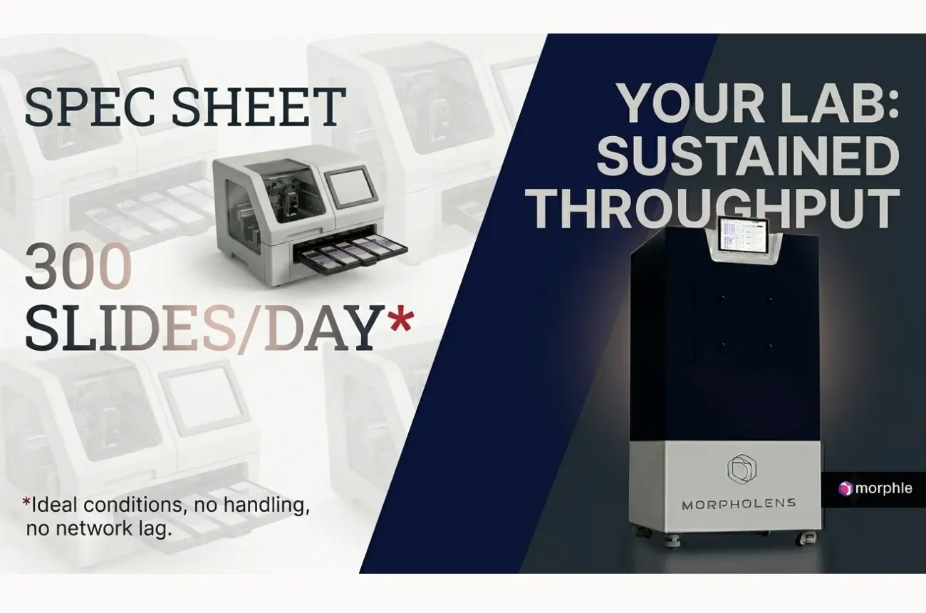
Digital slides are large; research institutions need efficient compression and storage planning. Systems like Morphle offer storage for approximately 3,000 scans on-board, simplifying dataset creation.
A lag-free experience is essential when dealing with high-magnification research imagery. A system that mimics microscope-like pan and zoom improves productivity and collaboration.
Unlimited user licenses ensure all lab members, collaborators, and students can access the dataset simultaneously.
Morphle scanners support research-driven digital pathology with consistent imaging, reliable focus, and accurate color calibration—key for reproducibility and quantitative analysis. With onboard storage for ~3,000 scans and unlimited user access, Morphle makes dataset creation and collaboration seamless. Flexible on-prem, cloud, or hybrid deployment and a responsive, microscope-like viewer ensure research teams can scale workflows and stay ready for AI-driven studies.
Digital pathology supports reproducibility—one of the biggest challenges in scientific research. Digital images remain consistent over time, enabling reanalysis, peer validation, and external review. Automated slide capture reduces human error and supports quantification for AI and computational pathology. Multi-user access accelerates teamwork and learning, particularly in large research programs or educational institutes.
Initial infrastructure costs, large data storage requirements, and the need for IT integration may be hurdles for some institutions. Additionally, research teams may require training as they transition from analog microscopy to digital workflows. While limitations exist, the long-term benefits greatly outweigh early adoption challenges.
Research institutes work with diverse datasets, from human tissue samples to animal research models. Compliance may involve institutional review boards, GLP guidelines, or data retention mandates. A Whole Slide Imaging Scanner must support secure storage, controlled access, audit logs, and metadata integrity to ensure traceability and reproducibility. DICOM compatibility is increasingly essential for long-term archival and interoperability.
Digital pathology is being used across:
Digital workflows allow hundreds of slides to be shared, analyzed, and compared without the physical limitations of glass storage or transport.
When selecting the best Digital Pathology Slide Scanner for their requirements,, research institutes should evaluate:
For compact, research-friendly scanning, see: Single slide scanner
The next generation of digital pathology will expand research capability further. AI-first scanners, automated region detection, multi-modal slide imaging, cloud-native datasets, and computational pathology frameworks will become standard. Collaborative platforms will enable global research teams to analyze datasets together, strengthening reproducibility and accelerating discovery.
Digital pathology is moving from a supporting tool to a central component of biomedical research infrastructure.
Transitioning from traditional microscopy to digital pathology scanners empowers research institutes with accuracy, reproducibility, and collaboration at scale. With features like unlimited user access, high-quality imaging, and flexible deployment, digital systems enable faster scientific discovery and unlock new possibilities in AI-driven analysis.
Contact the experts to see how your research attempts can benefit from Morphle scanners.

.webp)
.webp)
.webp)
.webp)
.webp)
.webp)